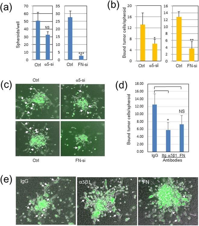
Figure 6.
Knockdown or antibody treatment of integrin (Itg) α5 in Panc-1 cells or fibronectin (FN) in WI-38 fibroblasts reduces fibroblast-dependent Panc-1 cell invasion in 3D collagen gel. Panc-1 cells and WI-38 cells were transfected with 10 nM control or specific siRNAs for Itg α5 or FN, respectively. These cells were harvested on the next day, mixed with non-treated counterparts, and incubated on an EZSPERE plate for 20 h. Chimeric spheroids thus produced were used in the following analyses. (a) Chimeric spheroid formation in α5-si Panc-1 (left panel) or FN-si WI-38 (right panel). Ordinate, the mean of the spheroid number per well ± SD in triplicate wells. NS, not significant (p = 0.056); ***p < 0.001. (b,c) Panc-1 cell invasion in contact with WI-38 cells in α5-si Panc-1 (left panel) or FN-si WI-38 (right panel). The above chimeric spheroids were incubated in collagen gel for 2 days, and Panc-1 cells elongated in contact with invading WI-38 cells, shown by arrows in (c), were counted for each spheroid. Ordinate, the mean of the number of elongate Panc-1 cells per spheroid ± SD in triplicate wells. The total spheroids analyzed were 14 in Ctr and α5-si Panc-1, 12 in Ctr WI-38, and 7 in FN-si WI-38. *p < 0.05, **p < 0.01. (c) Representative mages. Scale bars, 100 μm. See also Video 7. (d) Suppressive effects of anti-Itg-α5β1 and anti-FN antibodies on Panc-1 cell invasion. Invading Panc-1 cells in contact with fibroblasts were measured as above for each spheroid (total 6 to 8 spheroids per group). p = 0.066 in FN-si. (e) Representative images in (d).

