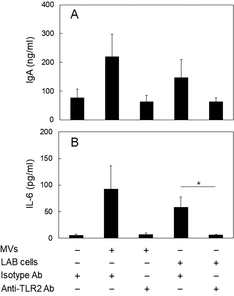
Fig. 4.
Involvement of TLR2 in the IgA-enhancing effects of the purified MVs and the cells of L. sakei NBRC15893.
Peyer’s patch (PP) cells (1.0 × 10⁵ cells/well) were cultured in the presence or absence of the anti-TLR2 antibody for 4 days with or without the MVs (protein concentration; 34 µg/ml) or L. sakei NBRC15893 cells (50 µg/ml). (A) IgA concentration and (B) IL-6 concentration in the culture supernatant was measured by ELISA. The data are expressed as means ± standard deviations of triplicate samples. *p<0.05, Student’s t-test.

