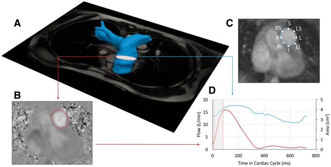
Figure 1.
(A) Magnetic resonance angiography reconstructed MPA with superimposed plane of phase-contrast CMR was applied to ensure the universally applied location for flow and stiffness analysis. (B) Exemplary phase-contrast image with the segmentation line derived from corresponding magnitude image. (C) Magnitude image with location specific labelled point of WSS analysis. (D) Created flow and area waveforms with highlighted region of the PWV analysis at the early ejection phase.

