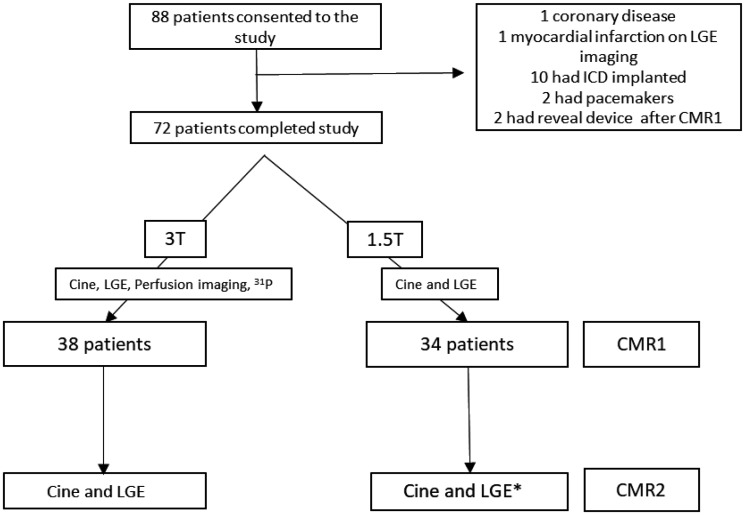
Figure 1.
A flowchart of hypertrophic cardiomyopathy (HCM) patients through the study. CMR, cardiovascular magnetic resonance imaging; ICD, implantable cardioverter defibrillator; LGE, late gadolinium imaging; 31P, Phosphorus-31 spectroscopy; T, Tesla. *CMR2 was at 1.5T or 3T (see Supplementary data online).

