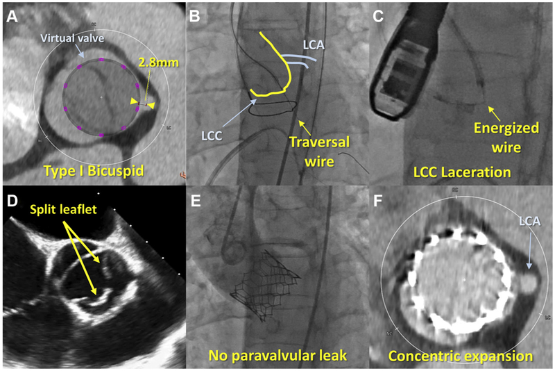
FIGURE 1. BI-SILICA During TAVR for BiAV.
(A) Sievers type-1 bicuspid morphology (fusion of left and right coronary cusps), with short distance of the virtual valve to coronary artery (2.8 mm). (B) An energized Astato XS20 guidewire traversed the left coronary cusp (LCC). (C) The energized guidewire lacerated the LCC. (D) Split leaflet after BI-SILCA was confirmed by transesophageal echocardiography. (E) Final aortography showed no paravalvular leak. (F) Post-procedural computer tomography revealed circular expansion of the balloon-expandable transcatheter heart valve and patent LCA. BiAV = bicuspid aortic valve; BI-SILICA = bicuspid scallop intentional laceration to induce circularization of the annulus; LCA = left coronary artery.

