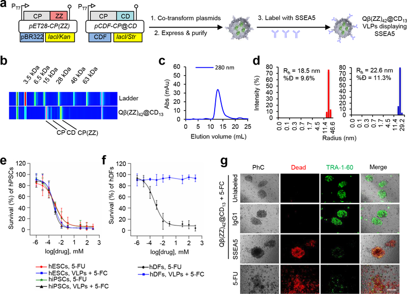
Figure 2. SSEA-5-labeled Qβ(ZZ)42@CD13 VLPs selectively kill hPSCs in the presence of 5-FC.
(a) Schematic of Qβ(ZZ)42@CD13 viral coat protein expression and particle assembly, followed by VLP labeling with SSEA-5 antibodies. (b) Electrophoretic, (c) FPLC, and (d) dynamic light scattering analyses of Qβ(ZZ)42@CD13 VLPs. (%Đ = % dispersity; Rh = hydrodynamic radius). (e) EC50 curves showing dose-dependent decreases in human induced pluripotent stem cell (hiPSC) and human embryonic stem cell (hESC) survival after 24 h treatment with both 8 nM Qβ(ZZ)42@CD13+SSEA-521 VLPs in the presence of the prodrug 5-fluorocytosine (5-FC) or the cytotoxin 5-fluorouracil (5-FU). (f) EC50 curves show dose-dependent decreases in human dermal fibroblast (hDF) survival percentages after 24 h treatment with 5-FU, but negligible cell death after treatment with 8 nM Qβ(ZZ)42@CD13+SSEA-521 VLPs in the presence of 5-FC. (g) Representative microscopy images show co-cultures of hPSC colonies and MEFs that were treated for 15 h with 8 nM of unlabeled, IgG1-labeled (isotype control), and SSEA-5-labeled Qβ(ZZ)42@CD13 VLPs in the presence of 100 μM 5-FC as test conditions, and 100 μM 5-FU as a positive control. Red fluorescence indicates dead cells and green fluorescence indicates expression of the pluripotent stem cell-specific TRA-1–60 marker.

