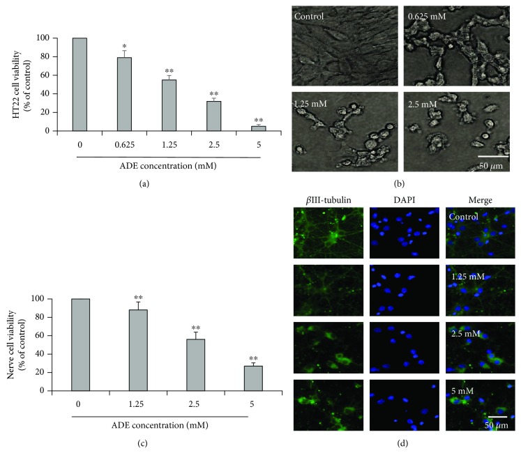
Figure 1.
Effect of ADE on the cell viability and apoptosis of HT22 cells. Cells were incubated with various concentrations of ADE for 24 h. (a) Cell viability of HT22 cells after treatment with ADE (0, 0.625, 1.25, 2.5, and 5 mM). (b) Cellular morphology of HT22 cells after treatment with ADE (0, 0.625, 1.25, and 2.5 mM). (c) Cell viability of primary cultured cortical neuronal cells after treatment with ADE (0, 1.25, 2.5, and 5 mM). (d) Cellular morphology of primary cultured cortical neuronal cells stained with βIII-tubulin and DAPI after treatment with ADE (0, 1.25, 2.5, and 5 mM) (n = 3, ∗p < 0.05, ∗∗p < 0.01 vs the control group). One-way ANOVA followed by Dunnett's multiple comparison tests was used to detect significant differences from the control.

