Figure 3.
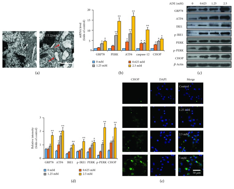
ADE induces ERS in HT22 cells. (a) Morphological changes of ER in HT22 cells after treatment with ADE were detected by TEM. The ER became swollen and vacuolated, as shown by red arrows. The white arrow indicates normal ER. (b) Effect of ADE on the mRNA levels of GRP78, PERK, ATF6, caspase-12, and CHOP in the HT22 cells. (c) After ADE treatment, the expression levels of GRP78, ATF6, IRE1, p-IRE1, PERK, p-PERK, and CHOP were elevated by western blot analysis. (d) ImageJ software was used to do the densitometric analysis of the ERS-related proteins. (e) Images of primary cultured cortical neuronal cells incubated with ADE for 24 h and stained with CHOP and DAPI. The targeted proteins were stained in green (CHOP), while the nuclei were stained in blue (DAPI) (n = 3, ∗p < 0.05, ∗∗p < 0.01 vs the control group). Statistical comparisons were made using one-way ANOVA with Dunnett's multiple comparison tests.

