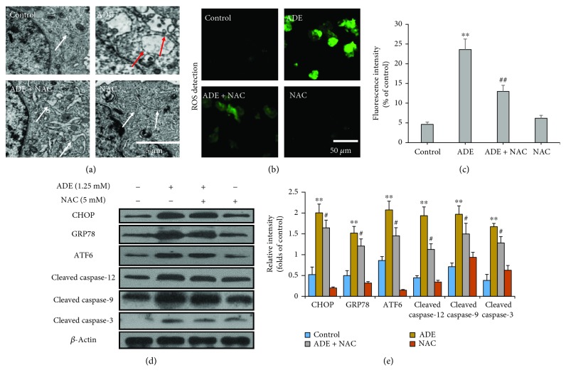
Figure 6.
Effect of NAC on ERS and apoptosis. (a) NAC markedly improved HT22 cells' morphological changes of ER after treatment with ADE. The red arrows indicate swollen and vacuolated ER, and the white arrows indicate normal ER. (b) Fluorescence images of NAC pretreatment cells stained by DCFH-DA (green) were obtained by confocal microscope. (c) Quantitative analysis of ROS-positive cell percentages in each group by ImageJ software (n = 3, ##p < 0.01, ADE+NAC vs ADE; ∗∗p < 0.01 vs the HT22 control group). (d) Western blot analysis showed that NAC markedly inhibited ADE-induced ERS- and apoptosis-related protein upregulation in HT22 cells. (e) Densitometric evaluation of western blot by ImageJ software (n = 3, #p < 0.05, ADE+4-PBA vs ADE; ∗∗p < 0.01 vs the HT22 control group). Statistical comparisons were made using one-way ANOVA with Dunnett's multiple comparison tests.

