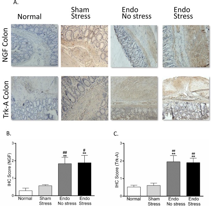
Figure 4.
Immunohistochemical staining in rat colon. (A) Representative pictures of NGF and Trk-A expression in colon (×40). Stress significantly increased the expression of (B) NGF and (C) its receptor Trk-A in colon (n = 6-8 animals per group [SEM], **P <.01 vs normal; # P <.05, ## P <.01 vs sham-stress). NGF indicates nerve growth factor; SEM, standard error of the mean.

