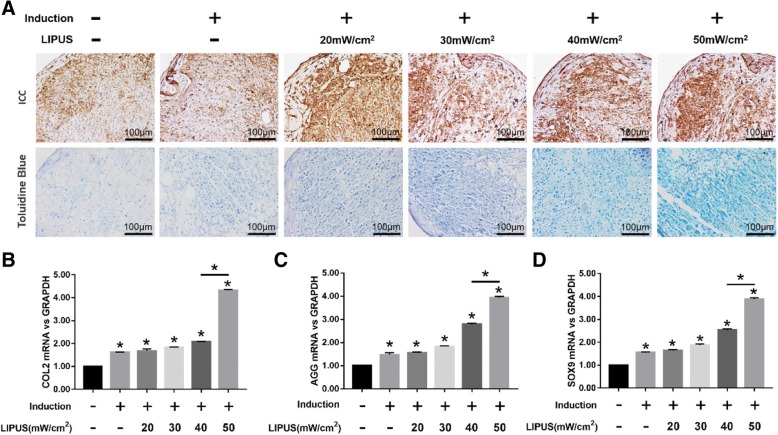
Fig. 3.
Effects of LIPUS on the chondrogenesis of MSCs. The MSCs were cultured in basic medium or chondrogenic medium for 10 days before analyses. a Representative images of ICC showing COL2+ cells (upper panel), and toluidine blue-staining showing AGG (lower panel) in differentiating MSCs stimulated with varying intensities of LIPUS; scale bars = 100 μm. b–d Bar graphs showing relative levels of COL2 (b), AGG (c), and SOX9 (d) mRNA in LIPUS-stimulated and unstimulated MSCs. The values are the mean ± SD of triplicate experiments; *P < 0.05

