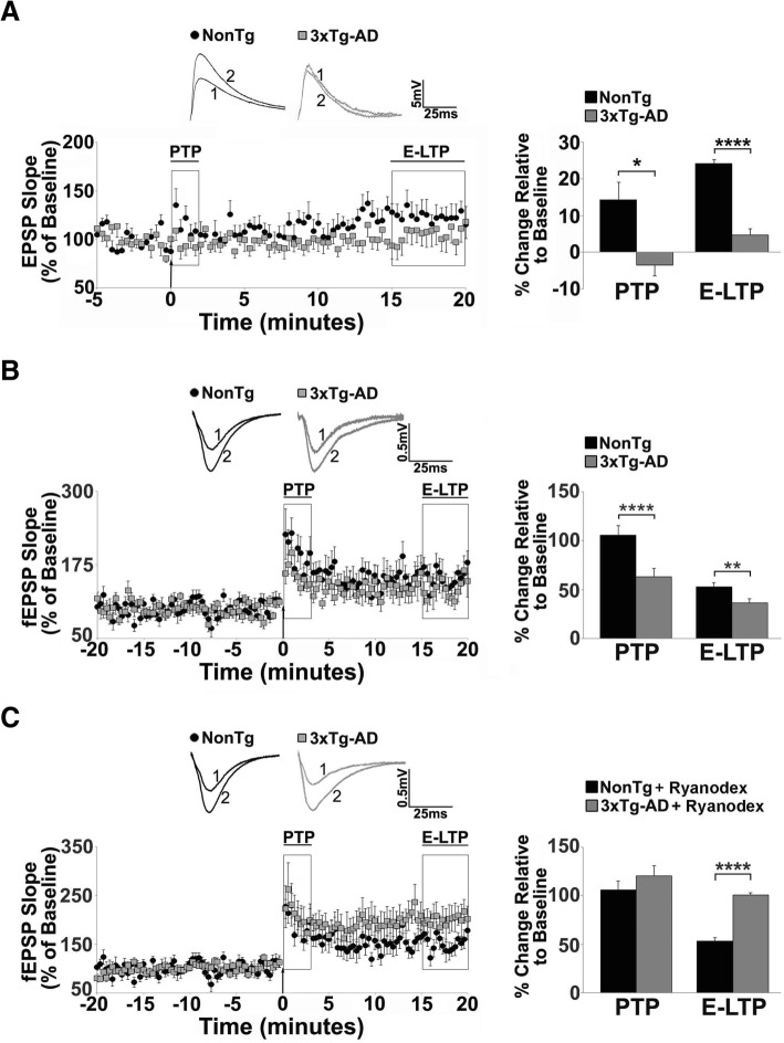
Fig. 1.
Short term plasticity deficits in 3xTg-AD CA1 region reflect RyR-Ca2+ abnormalities. a-c Left: Graphs show averaged time course of Schaffer collateral-evoked synaptic responses from NonTg (black circles) and 3xTg-AD (gray squares) in the hippocampal region using (a) whole cell patch clamp from CA1 pyramidal neurons (NonTg n = 10 neurons/6 mice; 3xTg-AD n = 10 neurons/6 mice) and (b-c) extracellular field potential recordings in CA1 stratum radiatum (NonTg n = 8 slices/5 mice; 3xTg-AD n = 8 slices/5 mice). Right: Bar graphs show averaged % change over baseline in the evoked response post-tetanus in 3xTg-AD (gray) compared to NonTg (black). Inset: Representative traces [1] before and [2] after tetanus from NonTg (black) and 3xTg-AD (gray) neurons. PTP and E-LTP are impaired in (a) individual neurons and (b) field potentials of 3xTg-AD CA1 neurons compared to NonTg neurons. c 30-day Ryanodex treatment restores or enhances PTP and E-LTP in 3xTg-AD CA1 circuits (n = 5 slices/5 mice; NonTg n = 5 slices/5 mice) . The arrow denotes the time of tetanus. Data are presented as Mean ± SEM; *p < 0.05, **p < 0.01 and ****p < 0.0001 represents significantly different from NonTg

