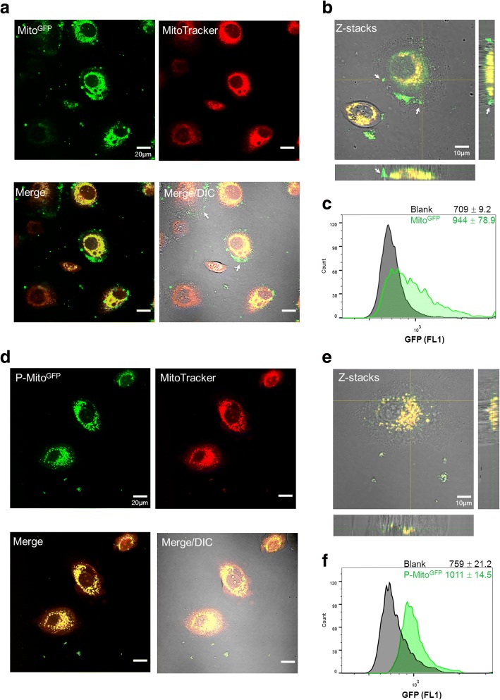
Fig. 1.
Expression of foreign mitochondria tagged with green fluorescent protein (MitoGFP) in MCF-7 human breast cancer cells pre-stained with MitoTracker Red. Internalization of MitoGFP (a-c) or Pep-1-labelled MitoGFP (P-MitoGFP) (d-f) was observed by confocal microscopy with different colour labels combined with the differential interference contrast (DIC)/bright field channel after 2-day treatments. The colocalization of foreign (green) and innate mitochondria (red) is shown in merged images (a, d) and Z-stacks (b, e), respectively. The white arrows indicate adhesion of Mito8344 to the outer cell membrane and entry failure (a, b). The quantification of mitochondrial internalization was performed by flow cytometry and is represented as the median fluorescence intensity of GFP with the standard deviation (c, f). Blank indicates the cell background of each group before treatment

