Figure 1.

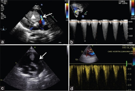
Transthoracic echocardiogram with color Doppler interrogation (a and b) of case 1 showing severe right ventricular outflow obstruction (arrow) by a large rhabdomyoma (solid triangle). (c and d) Regressed tumor with laminar flow

Transthoracic echocardiogram with color Doppler interrogation (a and b) of case 1 showing severe right ventricular outflow obstruction (arrow) by a large rhabdomyoma (solid triangle). (c and d) Regressed tumor with laminar flow