. 2019 Jan 17;114:e180332. doi: 10.1590/0074-02760180332

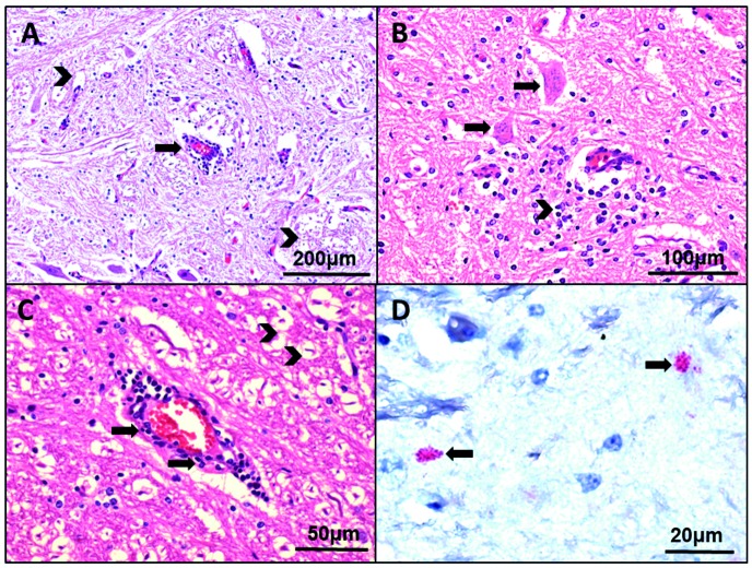
Fig. 2: photomicrograph of cerebral tissue of a horse with neurologic manifestation from EspĂ­rito Santo state with description of histopathological aspects and positive immunostaining for West Nile virus (WNV). (A) Inflammatory infiltrate predominantly constituted by mononuclear cells with perivascular cuffing (arrow) and neuronal necrosis (arrow heads). (B) Brain tissue showed neuronophagy (arrows) and associated gliosis (arrow head). (C) Perivascular infiltrate with congestion (arrows) and foci of neuronal necrosis of different intensities (arrow heads). (D) Immunohistochemistry of WNV antigen (reddish staining) in the cell cytoplasm in several foci of brain tissue (arrows).

Fig. 2: