Figure 1.
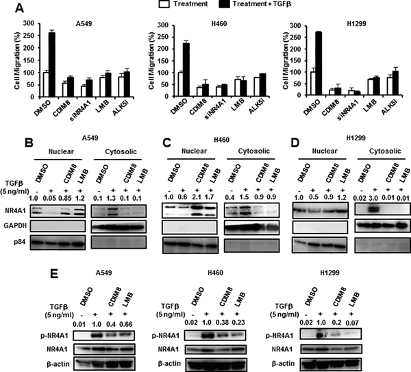
Role of NR4A1/CDIM8 in TGFβ-induced lung cancer cell migration. (A) A549, H460 and H1299 lung cancer cells were treated 5 ng/ml TGFβ (for 5 hr) and various reagents including siNR4A1 oligonucleotide (for NR4A1 knockdown), and cell migration was determined in a Boyden chamber assay. A549 (B), H460 (C) and H1229 (D) cells were treated with 5 ng/ml alone and in combination with LMB (20 nM) or CDIM8 (20 μM), and cytosolic and nuclear (B-D) or whole cell lysates (E) were analyzed by western blots. Results (A) are expressed as means ± SE for 3 separate determinations, and significant (p<0.05) induction of migration compared to solvent control (DMSO/CTL) is indicated (*). Bands in western blots (B-E) were quantitated relative to β-actin, and control values of NR4A1 were 1.0. The LMB and CDIM8 concentrations indicated above were used in subsequent experiments.

