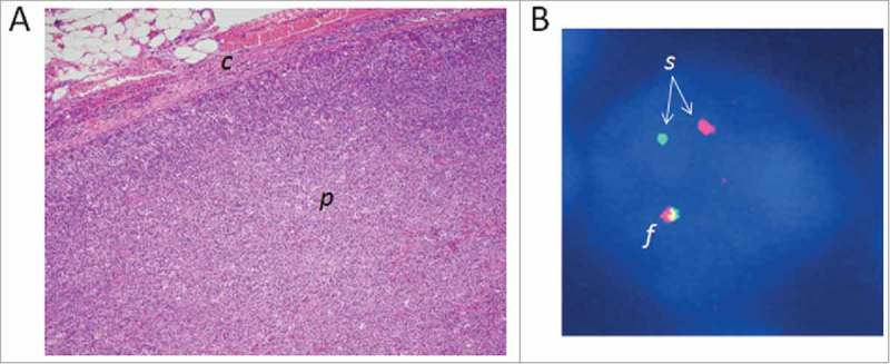
Figure 2.

Three-centimeter lymph node from a 55 year-old male with peripheral T-cell lymphoma, not otherwise specified, bearing a chromosomal rearrangement of the GEF gene VAV1. VAV1 rearrangements are seen recurrently in PTCL (see text).30 (A) Photomicrograph of the lymph node biopsy (hematoxylin and eosin stain; original magnification, 10×). The architecture of the lymph node parenchyma (p) has been effaced by the infiltrating lymphoma cells. c, lymph node capsule. (B) Fluorescence in situ hybridization (FISH) image of a single lymphoma cell nucleus (stained blue). The DNA has been hybridized with red and green fluorescent probes flanking the VAV1 locus. One VAV1 allele shows a normal red-green fusion signal (f), whereas the red and green signals of the other allele are split (s), indicating a VAV1 rearrangement.
