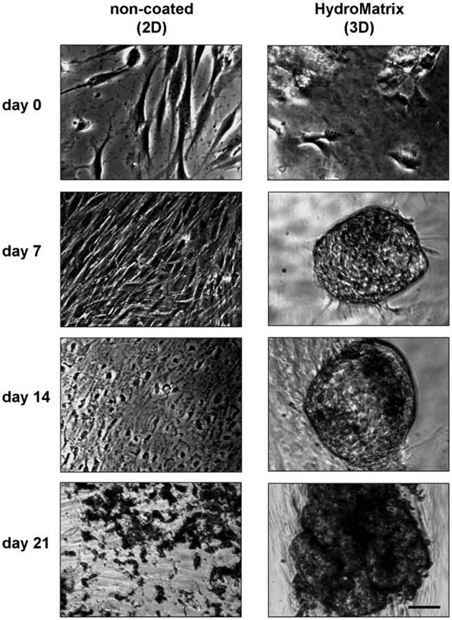
Fig. 4.

Morphological changes during osteogenic differentiation of PDLSCs on non-coated and HydroMatrix (HydM)-coated surfaces. Before osteogenic induction (day 0), low cell density and normal, fibroblast-like morphology can be observed both on plastic and gel surfaces. During the 3 weeks of the osteogenic differentiation, cell density increases. On plastic surface, the cells form multiple layers and significant amount of mineral deposits (dark spots) by day 21. In HydM, spherical cell clusters can be found from day 7 containing more and more mineralized matrix from day 14. Bars indicate 100 μm
