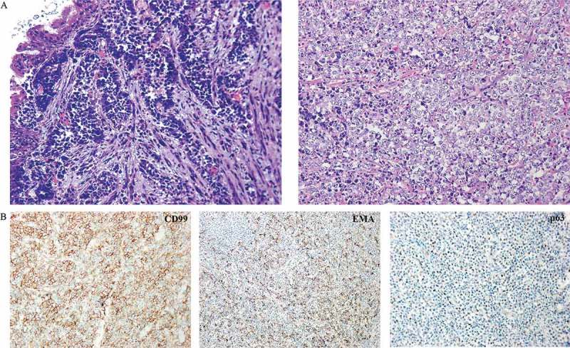
Figure 2.

Clinicopathologic characteristics of this case. (a) Pathology image of fiberoptic bronchoscopy deposits by hematoxylin and eosin stain showing round undifferentiated tumor cells. (b) Immunohistochemistry staining of tumor cells from a resected specimen that are positive for CD99, EMA and p63.
