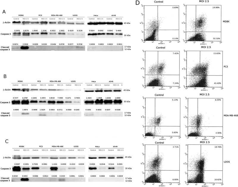
Figure 4.

CpHV-1 infection induces apoptosis in neoplastic cell lines. Panel A-C: Western blotting analysis of Caspase and Cleaved caspase 3 levels in MDBK, PC3, MDA-MB-468, U2OS, Hela and A549 cells were performed after mock infection or infection with CpHV-1 at MOI 2.5, at 12 h (panel A), 24 h (panel B) and 48 h (panel C) p.i.. β-Actin were used as a loading control. Band densitometry values indicate caspase 3 and cleaved caspase 3 levels normalized to β-Actin levels. Panel D: Annexin V apoptosis analysis. MDBK, PC3, MDA-MB-468 and U2Os cells were infected with CpHV-1 virus and after 24 h analyzed by annexin-V assay. The graphs (panel D) show the percentages of early apoptosis (population of cells positive for annexin-V staining) and late apoptosis/necrosis (population of cells positive for both annexin-V and propidium iodide staining).
