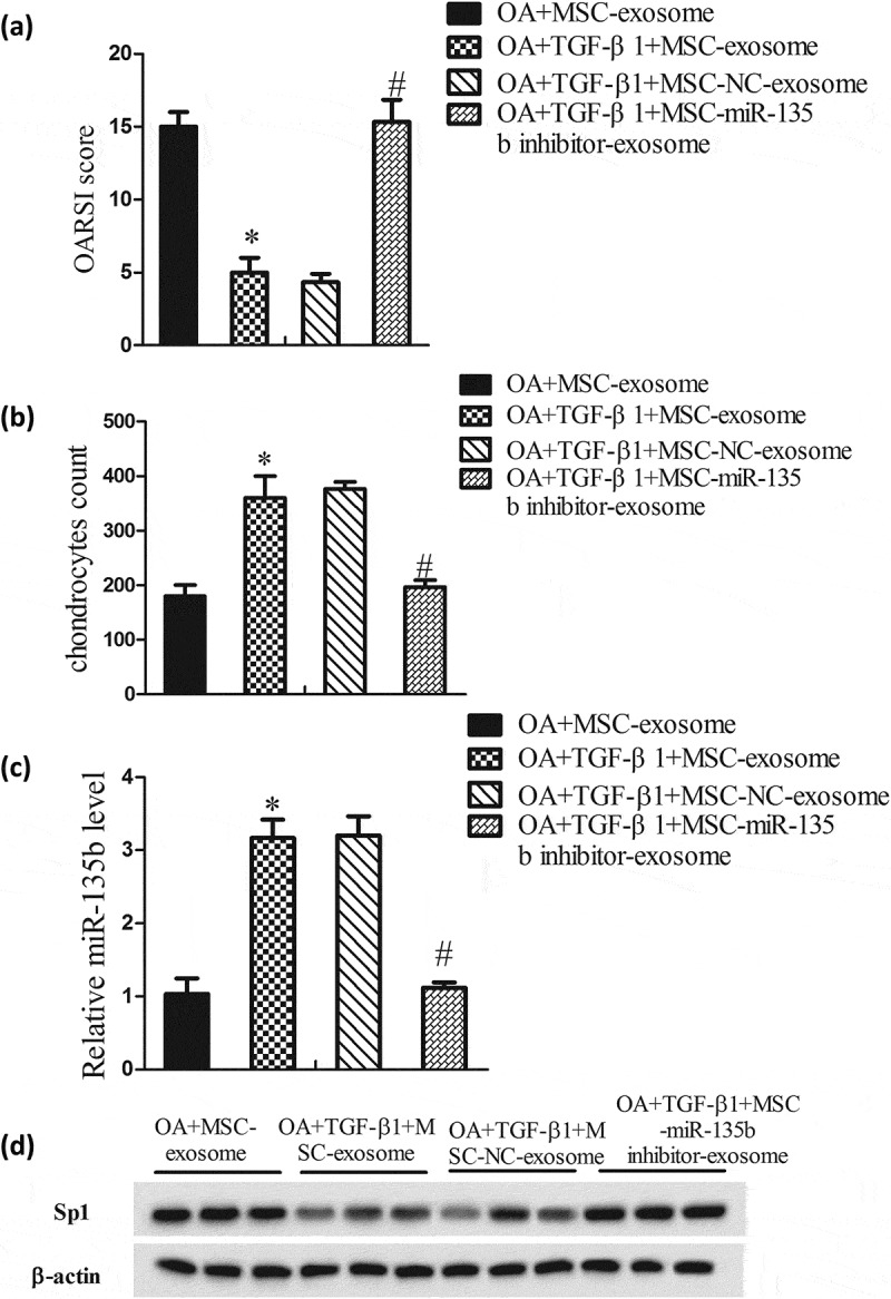
Figure 6.

TGF-β1 promoted the reparation of cartilage tissues in vivo. Rats were divided into OA+MSC-exosome group (rats received articular cavity injection of MSC-exosome, 100 μl; 1 × 10¹¹ MSC-exosome particles/ml; n = 6), OA+TGF-β1+ MSC-exosome group (rats received articular cavity injection of TGF-β1-exosome, 100 μl; 1 × 10¹¹ TGF-β1-exosome particles/ml; n = 6), OA+TGF-β1+ MSC-NC-exosome group (rats received articular cavity injection of TGF-β1-NC-exosome, 100 μl; 1 × 10¹¹ TGF-β1-NC-exosome particles/ml; n = 6), and OA+TGF-β1+ MSC-miR135b inhibitor-exosome group (rats received articular cavity injection of TGF-β1-miR135b inhibitor-exosome, 100 μl; 1 × 10¹¹ TGF-β1-miR135b inhibitor-exosome particles/ml; n = 6). Twelve weeks after surgery, rats were sacrificed and the knee samples were harvested to evaluate disease progression. (A) OARSI score. (B) Chondrocytes count. (C) MiR-135b expressions in cartilage. (D) Sp1 expression in cartilage. *P < 0.05, vs OA+MSC-exosome; #P < 0.05, vs OA+TGF-β1+ MSC-exosome.
