
Figure 2.
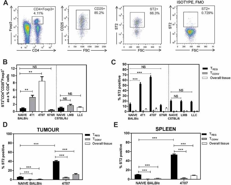
ST2+ Tregs accumulate in the lungs of mice bearing metastatic tumours 3 weeks post orthotopic implant. (a) Representative flow plots of lungs from a 4T07-bearing mouse; ST2 isotype fluorescence minus one (FMO) used for ST2 gating. (b) ST2+ Tregs, represented as a percentage of CD4+ T cells, are elevated in BALB/c mice bearing orthotopic 4T1 or 4T07 mammary carcinomas. Pulmonary ST2+ Tregs were not different in naïve vs EO771-LMB or LLC tumour-bearing C57BL/6 mice. (c) Elevated ST2+ Tregs in the lungs of 4T1 and 4T07 tumour-bearing mice. The percentage of Tregs expressing ST2 in the lungs was increased relative to ST2 expressing conventional CD4+ T cells (Tconv, CD4+CD25low/-Foxp3−). ST2+ Tregs were also increased in (d) primary tumour and (e) spleen of 4T07 tumour-bearing mice relative to naïve control tissue (mammary fat pads for tumours). Data are n = 4–6 mice per group from two independent experiments analyzed using Student’s two-tailed t-test, ** p ≤ 0.01, *** p ≤ 0.001.