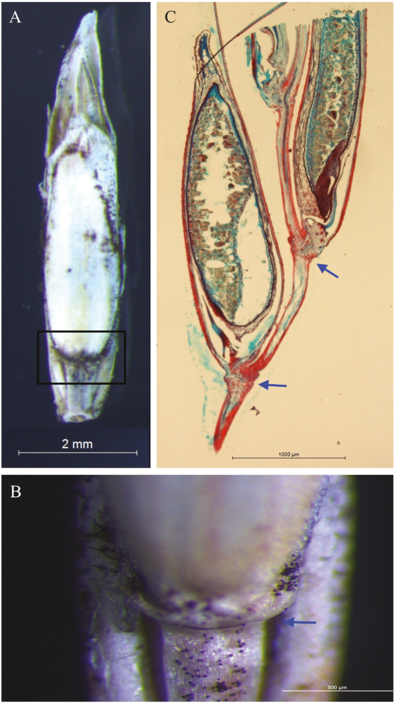
Figure 6.
Morphological and histological analysis of the abscission layer at the base of the seed within part of a spikelet. (A) Mature seed on spikelet of perennial ryegrass cv. Med line 1. (B) Enlarged images of the boxed area in (A), showing abscission layer located below the seed, indicated by the blue arrow. (C) Histological longitudinal sections of mature seed on a spikelet collected at 24 days after anthesis. Abscission layers are indicated by arrows.

