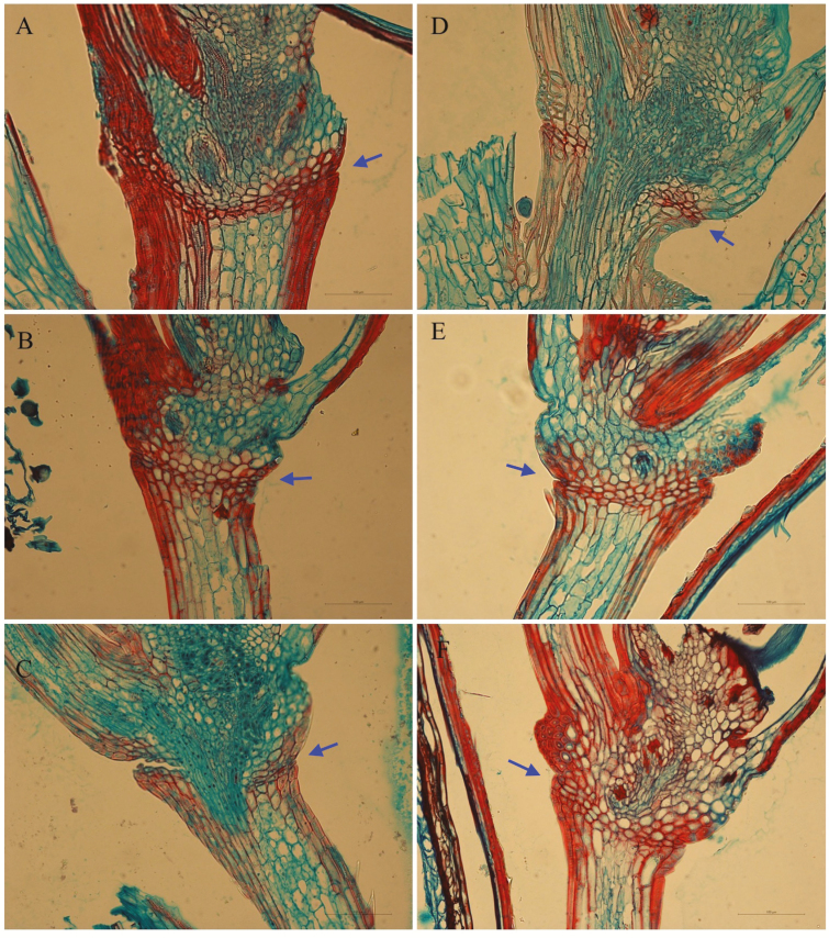
Figure 7.
Histological analysis of abscission layer. (A–F) Images of longitudinal sections across the abscission layer at 0 (A and D), 14 (B and E), 24 (C and F) days after anthesis for perennial ryegrass cv. Arrow (A–C) and cv. Med line 1 (D–F), respectively. Sections were stained with safranin-fast green, and the abscission layer is indicated by arrows.

