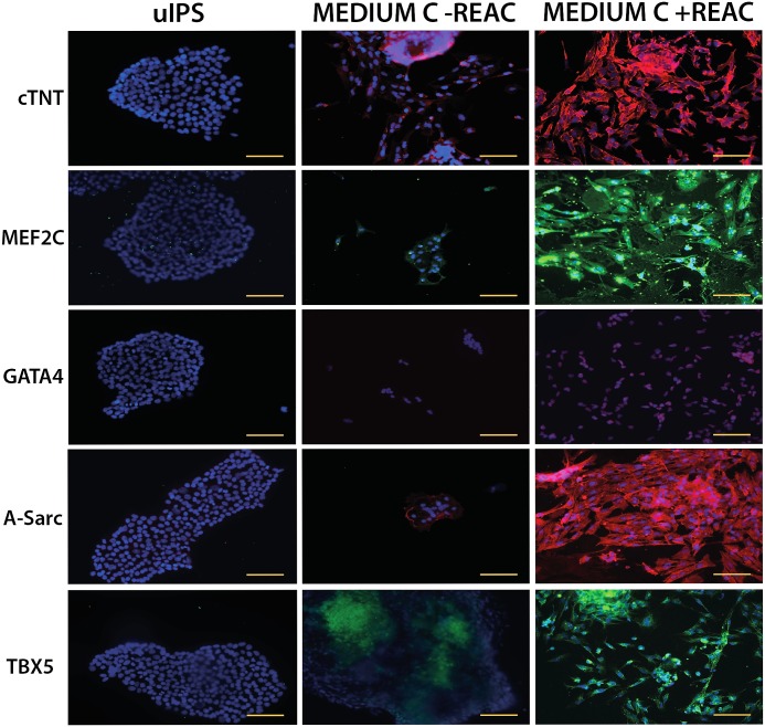
Fig 5. Immunohistochemical analysis of cardiac specific proteins.
hUiPSCs exposed for 72 hours to conditioned medium in the absence (Medium C-REAC) or presence of REAC (Medium C+REAC) were disaggregated after 14 days from time 0 (undifferentiated hUiPSCs) seeded in chamber slide and processed for immunostaining using specific antibodies directed against: Troponin complex (cTnT), MADS box transcription enhancer factor 2 (MEF2C), GATA binding protein 4 (GATA4), α-sarcomeric actinin (A-Sarc) and T-Box protein 5 (TBX5). Confocal microscopy analysis was performed with Leica confocal microscope (LEICA TCSSP5). Nuclei were labeled with DAPI (blue). The yellow scale bars are 100 μm.

