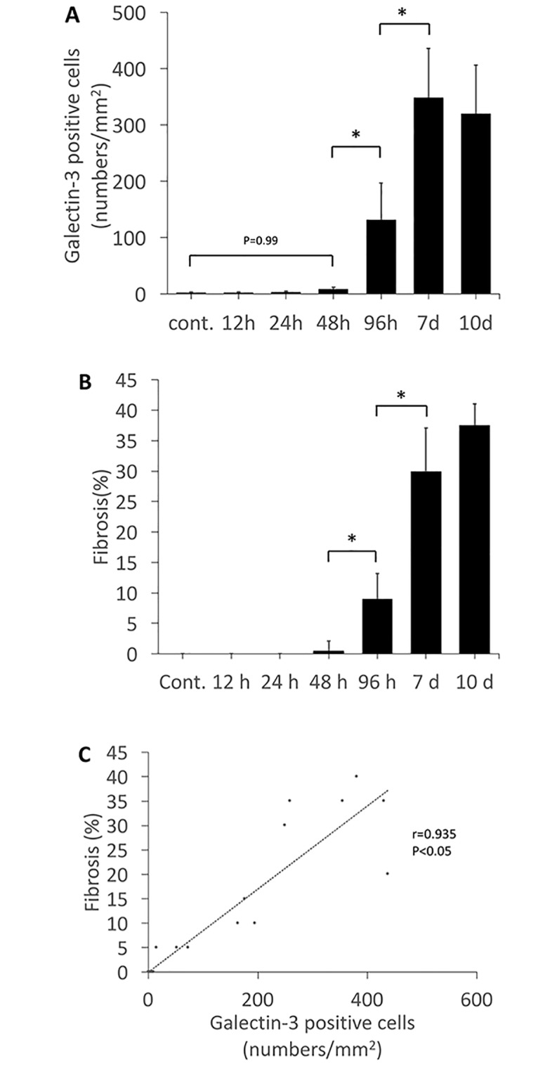
Fig 3. Quantification and association between the galectin-3 positive cell number and the degree of fibrosis.

A, The number of galectin-3 positive cells (determined by immunohistochemistry) showing myocardial infiltration was counted at each time point. B, The degree of myocardial fibrosis (determined by of Azan staining) was quantified as percentage of fibrotic area to total myocardial tissue at each time point. C, Association between galectin-3 positive cell number and degree of fibrosis. The galectin-3 positive cell numbers were positively correlated with the degree of myocardial fibrosis. *Statistically different from each group (p < 0.05), determined by ANOVA.
