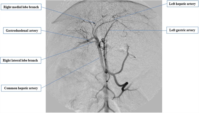
Fig 2. Ventral-dorsal digital subtraction images of a canine.
Hepatic arteriogram was performed after the injection of contrast medium through a 4-French catheter placed in the common hepatic artery. We inserted a catheter in the left hepatic artery, right medial lobe branch, and right lateral lobe branch and injected bone marrow-derived mesenchymal stem cells (BMSCs) via each of these arteries at doses of 2 × 10⁵ cell/kg, 1 × 10⁵ cell/kg, and 1 × 10⁵ cell/kg, respectively.

