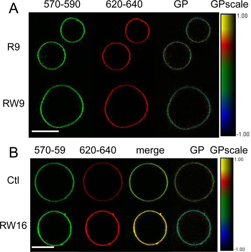
Fig 6. Confocal images of GPMVs labelled with di-4-ANEPPDHQ and incubated with CPPs.
The 570-590 nm range corresponds to the ordered membrane contribution and the 620-640 nm range to the disordered membrane contribution. GP was calculated as explained in the methods section. (A) GPMVs incubated with R9 (top) and RW9 (bottom) during 190 min. (B) Peptide-free GPMV (top) and incubated with RW16 (bottom) during 60 min. Bars represent 10 μm.

