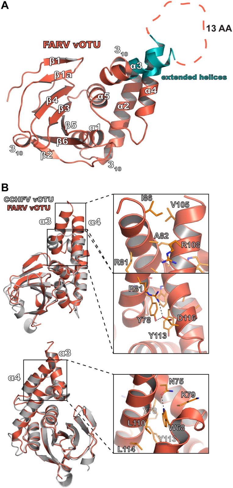
Fig 6. Structure of the FARV vOTU.

(A) Overall structure of the FARV vOTU with the secondary structure denoted based on DSSP. The extended regions of the α3 and α4 helices are colored in teal. Intervening amino acids lacking electron density are represented by an orange dashed line. (B) Molecular features of the extended α3-α4 helices of FARV vOTU, with CCHFV vOTU included for comparison. Atom pairs within hydrogen bonding distance are denoted by black dashes.
