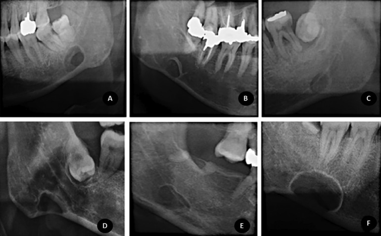
Figure 1.
Examples of bone margin and locularity classifications, from left to right: Figure 1A demonstrates a multilocular defect with total thin sclerosis; 1B demonstrates a unilocular defect with thick sclerosis; 1C demonstrates a unilocular defect without sclerosis at bone margins. Examples of the topographic relationship between the mandibular canal and the defect: Figure 1D shows a defect below the mandibular canal; Figure 1E shows a defect overlapping the mandibular canal inferior wall and below the mandibular canal upper wall; Figure 1F exhibits a defect that overlaps the upper and inferior walls of the mandibular canal.

