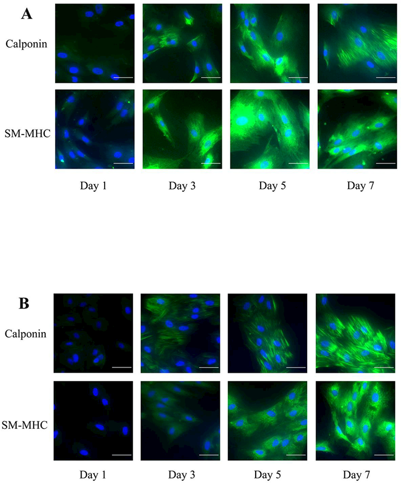
Figure 4.

The immunofluorescence staining of SMC-specific marker calponin and SM-MHC over 7 day differentiation from (A) BMSCs and (B) ADSCs. Nucleus were stained with DAPI while antibodies against calponin and SM-MHC were used for immunostaining (scale bar=50μm). Note that some of the ADSC cells in the day 3 images do not show calponin staining.
