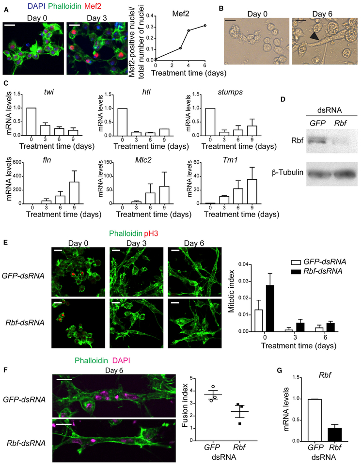
Figure 1. The Function of Rbf Is Required to Differentiate Dmd8 Cells into Myotubes.
(A) Dmd8 myoblasts were treated with differentiation medium for 0, 2, 4, and 6 days and stained with anti-Mef2 antibody, phalloidin and DAPI. Confocal images at day 0 and 3. Quantification of the number of Mef2-positive nuclei normalized to the number of nuclei. Mean. Experiment was done at least twice with n = 10 images with 80–400 cells per image.
(B) Brightfield image of elongated myotube at day 6 after treatment.
(C) mRNA expression level of the genes twist (twi), heartless (htl), stumps, flightin (fln), Myosin light chain 2 (Mlc2), and Tropomyosin 1 (Tm1-H) at day 0,3,6, and 9 after treatment. Mean ± SEM, n = 2.
(D) Lysates of Dmd8 cells treated for 4 days with GFP-dsRNA and Rbf-dsRNA, blotted against Rbf and β-tubulin.
(E) Confocal images of Dmd8 cells treated with GFP-dsRNA and Rbf-dsRNA at day 0, 3, and 6, stained with phalloidin and anti-pH3. Quantification of the number of pH3-positive nuclei normalized to number of nuclei. Mean ± SEM, experiment was done three times once with n = 6 images and twice with n = 10 images with 100–400 cells per image, two-way ANOVA, p > 0.05.
(F) Confocal images of Dmd8 cells treated with GFP-dsRNA and Rbf-dsRNA at day 6, stained with phalloidin and DAPI. Quantification of the number of nuclei per myotube. Mean ± SEM, experiment was done three times with n = 10 images with between 2 and 6 myotubes per image, Mann-Whitney test, p > 0.05.
(G) mRNA expression levels of Rbf at day 6 after treatment. Mean ± SEM, n = 2. Mann-Whitney test, p > 0.05.
Scale bar (A, B, E, F), 10 μm.

