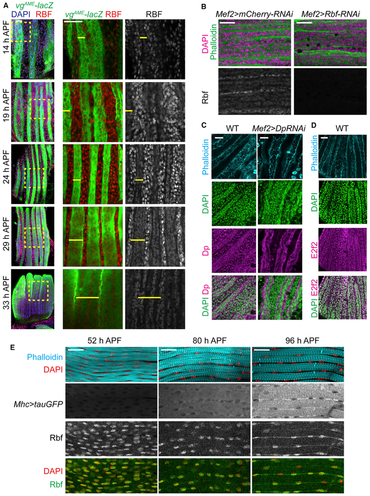
Figure 6. The Expression Level of Rbf Switched from Proliferating Myoblasts to Developing Myotubes.
(A) Developing DLMs labeled using vgAME-lacZ reporter (yellow bar) and anti-Rbf antibodies from 14 to 33 h APF. Magnified images of the yellow, dashed boxes indicated on the overlay image on the left.
(B) Developing DLMs at 29 h APF stained with anti-Rbf antibody, phalloidin, and DAPI in Mef2>mCherry-RNAi and Mef2>Rbf-RNAi as negative control.
(C and D) Developing DLMs staged at 24 h APF (C) and 21 h APF (D) stained with phalloidin, DAPI, anti-Dp (C) and anti-E2f2 (D) antibodies. Mef2>Dp-RNAi was used as negative control.
(E) Developing DLMs labeled using Mhc>tauGFP, anti-Rbf antibody, phalloidin, and DAPI from 52 to 96 h APF.
Genotypes are (A) w-;vgAME-lacZ, (B) w-, UAS-Dicer2; +; Mef2-GAL4/UAS-mCherry-RNAi, and w-, UAS-Dicer2; +; Mef2-GAL4/UAS-Rbf-RNAi, (C) w-;vgAME-lacZ as WT and w-; UAS-Dp-RNAi; Mef2-GAL4, (D) y-w-, and (E) w-; P{Mhc-tauGFP}2. Scale bars, 20 μm.

