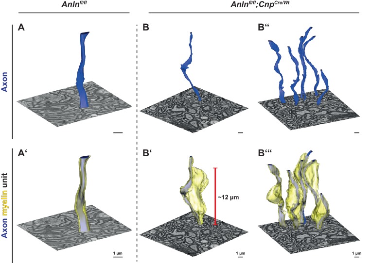
Figure 4. Three-dimensional reconstruction of myelin outfoldings in Anln cKO mice.
(A–C) Focused ion beam-scanning electron microscopy (FIB-SEM) micrographs and 3D reconstruction of the plasma membrane of myelinated axonal segments (blue) and respective myelin sheaths (yellow) of representative axon/myelin-units in the optic nerve of control (Anlnfl/fl) (A,A‘) and Anln cKO (B,B‘,B‘‘,B‘‘‘) mice at 5.5 mo. Note the tight association of the myelin sheath reconstructed in A ‘with the corresponding axon (in A,A‘) over at least 10 µm in the control nerve. An individual myelin outfolding (B‘) and the corresponding axon are reconstructed over 20 µm in B,B‘. All myelin outfoldings in that same block (as in B,B‘) were reconstructed in B‘‘‘ with their corresponding axons (in B‘‘,B‘‘‘). Note that myelin outfoldings represent large sheets of compacted multilayered membrane stacks that extend considerably away from their respective myelinated axon, commonly displaying longitudinal dimensions between 10 µm and 15 µm. See Videos 1–3.

