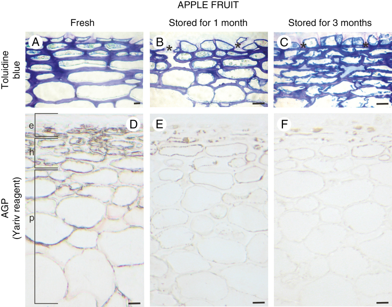
Fig. 1.
Histology of fruit tissue during senescence and its correlation with the presence of AGP. Toluidine blue-stained sections of apple fruit: fresh (A), after 1 month of storage (B), and after 3 months of storage (C). The deep apertures in the epidermal layer are asterisked in B and C. Red stain confirming the presence of AGPs in apple fruit: fresh (D), stored for 1 month (E), and stored for 3 months (F), after staining with Yariv reagent. Light microscopy. Scale bars = 10 µm (A–C), 20 µm (D–F). Abbreviations: e – epidermis, h – hypodermis, p – parenchymal cells.

