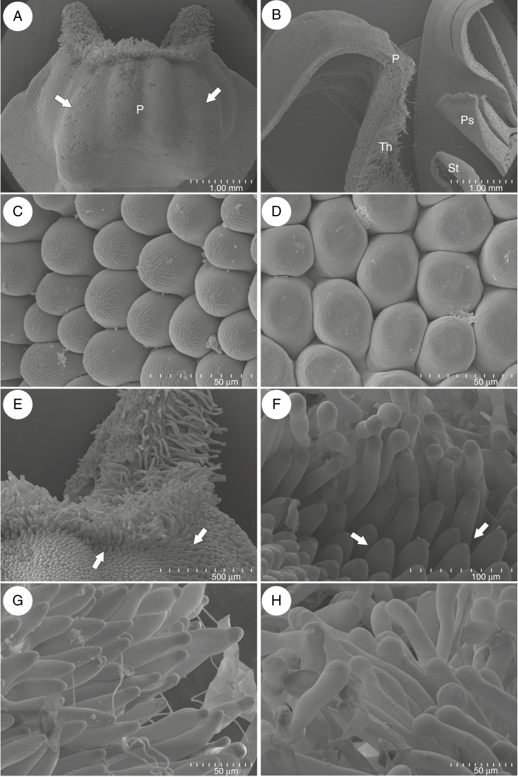
Fig. 2.
Micromorphology of Utricularia menziesii lower corolla lip. (A) Base of lower corolla lip with palate (P); note the clearly visible parallel ridges; the inner ridges were smaller than the outer ones (arrows); scale bar = 1 mm. (B) A part of a section through the flower showing: palate (P), throat (Th), pistil (Ps) and stamen (St); scale bar = 1 mm. (C). Micromorphology of epidermal cells of palate, note cuticular striations; scale bar = 50 μm. (D) Micromorphology of epidermal cells of the lower lip; scale bar = 50 μm. (E and D) Micromorphology of palate base, note papillae (arrows) and trichomes; scale bar = 500 μm and scale bar = 100 μm. (G–H) Trichomes of palate; scale bar = 50 μm.

