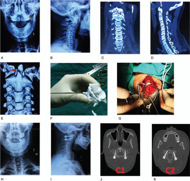
Figure 2.

A 45-year-old man suffered traffic accident injury. (A, B) Preoperative anteroposterior and lateral X-ray view of cervical spine showed C2 odontoid fracture. (C–E) Preoperative coronal position, sagittal position, and 3-dimensional (3D) reconstruction image of computed tomography (CT) scans also showed C2 odontoid fracture. (F) Simulation operation in the printing navigation model preoperatively. (G) Placing the 3D printing model intraoperatively. (H, I) Postoperative cervical anteroposterior and lateral X-ray showed the internal fixation is well positioned. (J, K) Postoperative CT scans confirmed the pedicel screws were safely inserted into the C1 and C2 pedicles.
