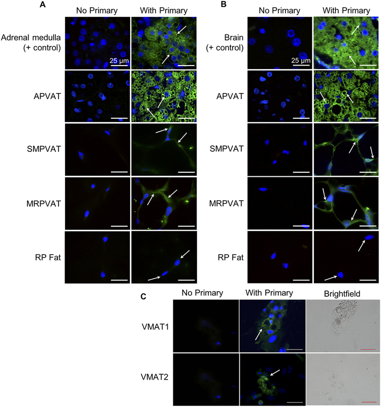
Figure 3.
VMAT1 and VMAT2 are present in aortic (A, 2nd rows), superior mesenteric (SM, 3rd rows) and mesenteric (MR, 4th rows) PVATs, but not retroperitoneal fat (RP fat, bottom rows). A and B.
Immunofluorescence images using a primary antibody against VMAT1 (A) or VMAT2 (B) on right and negative control image for each tissue on left. Images were taken with a 100× oil objective. Arrows indicate areas of interest/positive staining (green). All scale bars represent 25 μm. Representative of 4–6 animals. Positive control tissues (top rows) were rat adrenal medulla (A) and the anterior commissure of the rat brain (B). VMAT1 and VMAT2 staining is present in adipocytes differentiated from adipocyte precursors in MRPVAT SVF (C). Scale bars represent 50 μm. Representative of 3 animals.

