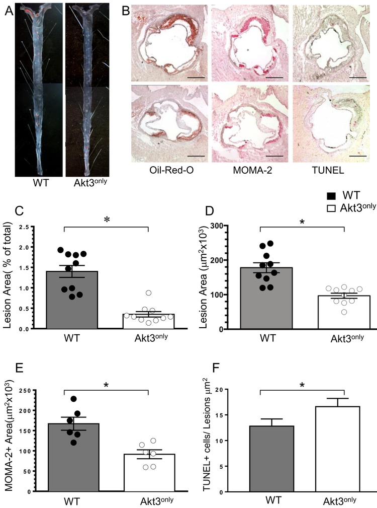
Figure 3. Reconstitution of Akt3only hematopoietic cells into male Ldlr−/− mice reduces atherosclerosis.
(A) Detection of atherosclerotic lesions in pinned out aorta en face of WT→Ldlr−/− and Akt3only→Ldlr−/− mice; Aortas were stained with Sudan IV, pin size 10μm.
(B) Detection of atherosclerotic lesions, MOMA-2+ area and apoptotic cells in aortic sinus of WT→Ldlr−/− (top) and Akt3only→Ldlr−/− mice (bottom); Aortic sections were stained with Oil-Red-0, MOMA-2, or TUNEL assay; Scale bars, 200μm.
(C-F). The percent of atherosclerotic lesions in pinned out aortas (C), the extent of lesion area (D), MOMA-2+ area (E) and number of TUNEL+ cells (F) in aortic sinus of male Ldlr−/− mice reconstituted with WT or Akt3 FLC; Graphs represent data (mean ± SEM, ∗p<0.05 by Mann-Whitney Rank Sum and *p<0.05 by t-tests).

