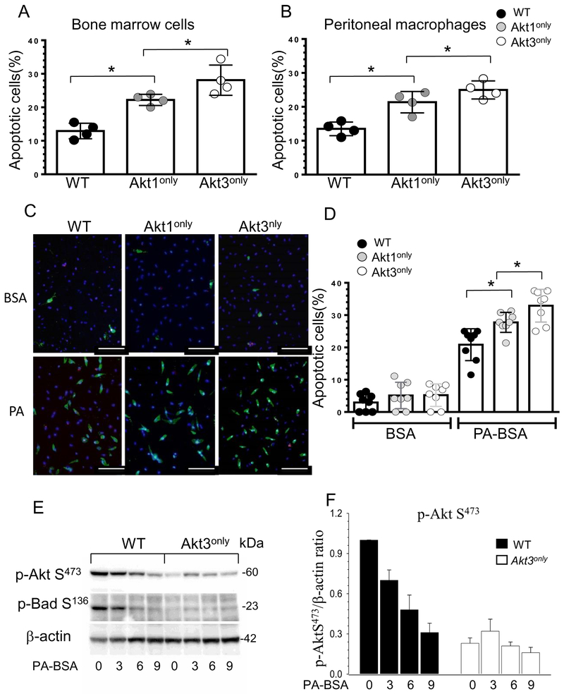
Figure 5. Blood Akt1only or Akt3only monocytes are more sensitive to ER stress than WT cells(A,B).
Apoptotic cells in bone marrow and peritoneal macrophages freshly isolated from Ldlr−/− mice reconstituted with WT, Akt1only and Akt3only FLC; cells were incubated with Alexa Fluor 647 Annexin V antibody and analyzed by flow cytometry; *p<0.05 compared to WT cells by One Way Analysis of Variance (Tukey test).
(C,D) Detection and the percent of apoptotic monocytes after treatment with BSA or PA-BSA overnight. Monocytes were isolated from blood of Ldlr−/− mice reconstituted with Akt1only, Akt3only or WT FLC, seeded in culture chambers and, two days later, treated with BSA or a pro-apoptotic stress factor, PA-BSA in the presence of 3% fetal bovine serum and 10nM of mouse macrophage colony-stimulating factor overnight, then apoptotic cell numbers were analyzed using Alexa Flour 488 Annexin V/Dead cell apoptosis kit. Graphs represent data (mean ± SEM) obtained from the same (n=8/group) mice; *p<0.05 compared to WT cells treated with PA by One Way Analysis of Variance, Dunnett’s method); Scale bar is 50mm.
(E,F) ER stress inhibits Akt signaling faster in Akt3only macrophages than in WT cells. WT, Akt3only peritoneal macrophages were treated with BSA or 0.5M PA-BSA for 0, 1, 3 and 6 hours. Extracted proteins were used for analysis of Akt and BadS136 signaling. Graphs represent data (mean ± SEM) of three experiments.

