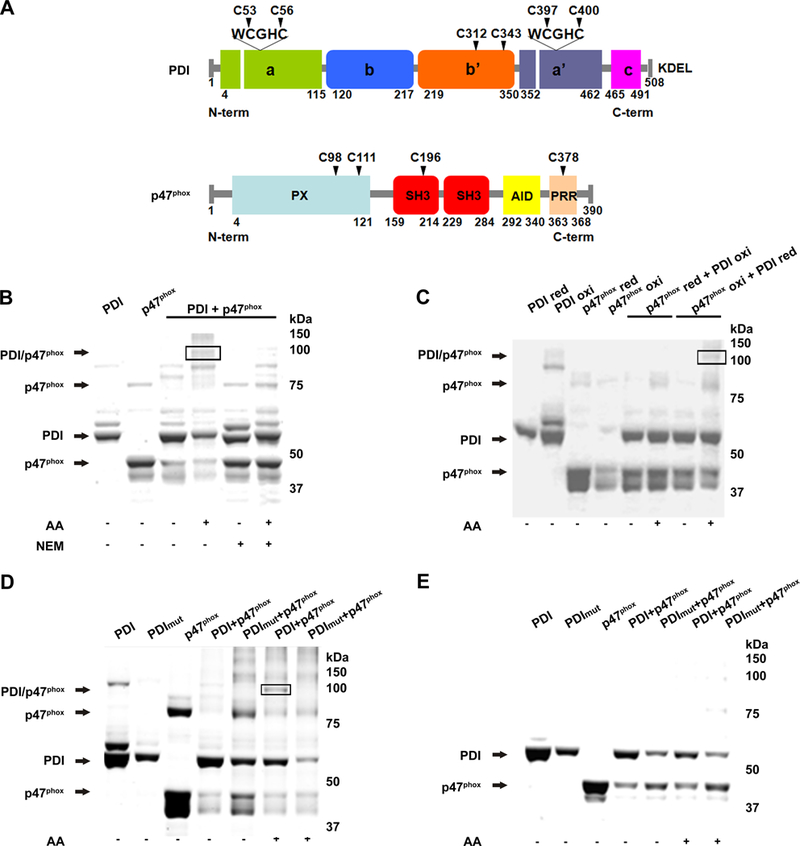
Figure 1. PDI interacts with p47phox through its redox active sites.

(A) Cysteine positions within domains of PDI and p47phox. (B) Non-reducing polyacrylamide gel stained with Coomassie blue shows monomers and dimers following incubation of recombinant wt PDI and p47phox with and without NEM. (C) Combinations of reduced PDI (PDI-red) and oxidized p47phox (p47phox-oxi), and oxidized PDI and reduced p47phox were analyzed as in B. (D) PDI mutated at the four redox cysteines (PDI mut) was reacted with p47phox and resolved in non-reducing (D) and reducing (E) polyacrylamide gels. AA: arachidonic acid. n=2.
