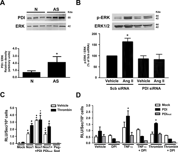
Figure 2. PDI is increased in atherosclerosis and regulates Nox1 NADPH oxidase-mediated signaling.

(A) Expression of PDI in non-human primate aorta (N: Normal, AS: atherosclerotic) n=3, *p<0.05 vs N. Data normalized to total ERK2 levels. (B) Ang II-induced ERK 1/2 phosphorylation after PDI silencing in VSMC. Quantification normalized to total ERK 1/2 levels. n = 3, *p<0.05 vs scr. (C) Superoxide levels in HEK-293 cells after transfection with PDI or PDI mut treated with DPI, n=3, *p<0.05 vs mock. (D) Superoxide levels measured by L-012 chemiluminescence (RLU) in Nox1-/y VSMC after transfection with Nox1, PDI or PDI mut and stimulated with thrombin. *p<0.05 vs Mock vehicle, # p<0.05 vs Mock thrombin, • p<0.05 vs Nox1 vehicle, ◦ p<0.05 vs Nox1/PDI vehicle, + p<0.05 vs PDI thrombin, n=3.
