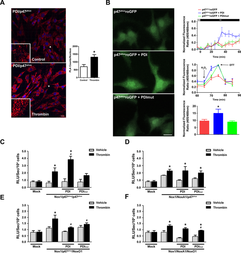
Figure 3. PDI increases Nox1 activation through a redox dependent interaction with p47phox.

(A) Representative Z-stack images of Duolink analyses of PDI interaction with p47phox in VSMC stimulated or not with Thrombin (Imaris, Bitplane, Version 7.6.5). Positive signals demonstrating an interaction of the indicated proteins are shown as red dots, DAPI (blue), n=3. 41 slices were quantified using ImageJ. *p< 0.05 vs Control (B) Representative time course of p47-roGFP in rabbit VSMC. Cells were dually excited with 405 and 488 laser lines and the emitted fluorescence at 505–550 nm was captured at 20s sampling intervals. Representative data of at least 15 cells for each condition. * p< 0.05 vs p47-roGFP. (C-F) COS p22 phox cells were transfected with indicated subunits and superoxide levels measured by L-012 chemiluminescence (RLU). n=6 for each condition, * p< 0.05 vs mock, # p< 0.05 vs thrombin with no PDI.
