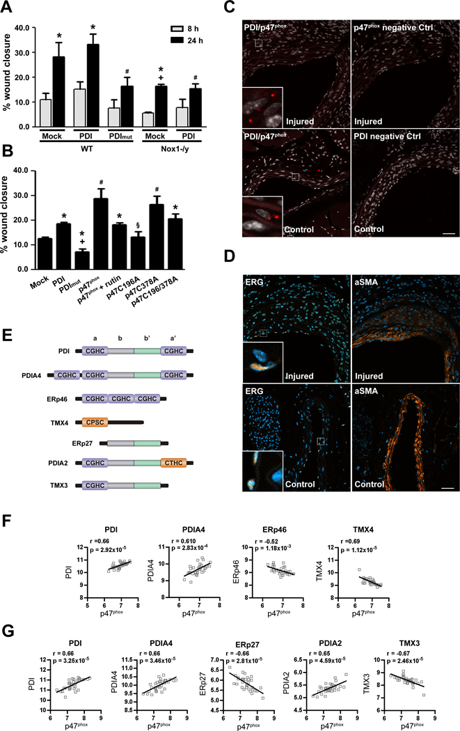
Figure 6. p47phox interacts with PDI in vivo.

(A) VSMC migration to thrombin is dependent on Nox1 (n=3 * p<0.05 vs 8h, # p<0.05 vs PDI 24h in WT and + vs mock 24h). (B) Rabbit aortic VSMC were transfected as shown and 48 hours later stimulated with angiotensin II for 8 hours. In some experiments, rutin (100 μM), a PDI-inhibitor, was added prior to angiotensin II. (n=5, * p<0.05 vs p47phox wt, #p<0.05 vs mock, + p<0.05 vs PDI, § p<0.05 vs p47phox, p47phox C378A). (C) Interaction of PDI and p47phox after carotid artery wire-injury, postoperative day 14. Representative images of Duolink analyses of the interaction of PDI and p47phox and the respective controls as indicated. Positive signals demonstrating an interaction of the indicated proteins are shown as red dots. Co-staining with DAPI (white) to show the nucleus. Auto fluorescence shows the elastic lamina (red). The scale bars represent 40μm. n= 3. (D) Representative immunofluorescence images of carotid artery wire-injury, postoperative day 14. Staining against endothelial cell marker ERG (brown) and alpha-smooth muscle actin (α-SMA, brown) are shown as indicated. Co-staining with DAPI (blue) to show the nucleus. (E) PDI family of thiol isomerases structures. Pearson correlation analysis of p47phox and PDI family members using mRNA expression levels from 32 paired samples of macroscopically intact tissue (F) and atheroma plaque (G) was retrieved from Gene Expression Omnibus (GEO, GSE43292).
