Figure 1.

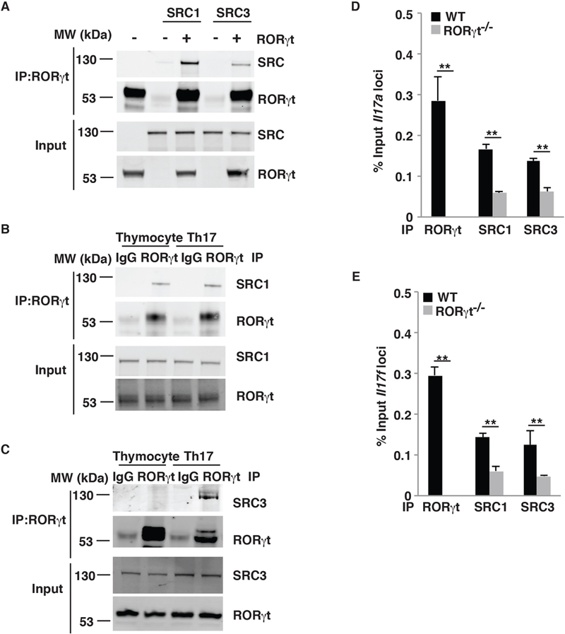
SRC3 interacts with RORγt in Th17 cells but in not thymocytes. (A) Immunoblot analysis of SRC1 and SRC3 immunoprecipitated with an anti-RORγt antibody from HEK293T cells co-transfected with plasmids to express RORγt, SRC1, and/or SRC3. The bottom images show immunoblot analysis of whole-cell lysates without immunoprecipitation (input). (B) Immunoblot analysis of SRC1 immunoprecipitated with a control IgG or anti-RORγt antibody from mouse thymocytes and differentiated Th17 cells. (C) Immunoblot analysis of SRC3 immunoprecipitated with a control IgG or anti-RORγt antibody from mouse thymocytes or differentiated Th17 cells. (D) ChIP analysis of RORγt, SRC1, and SRC3 bound to the Il17a locus in CD4+ T cells from WT or RORγt−/− mice polarized under Th17 conditions. (E) ChIP analysis of RORγt, SRC1, and SRC3 bound to the Il17f locus in CD4+ T cells WT or RORγt−/− mice polarized under Th17 conditions. Data are from one representative of three independent experiments (A–C) or are aggregated from three experiments (D–E, mean ± SEM). ** P <0.01 (t-test).
