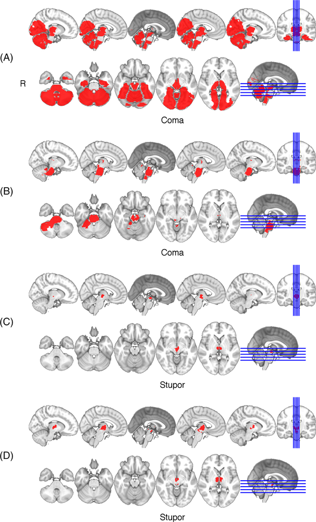
FIGURE 2:
Lesions in patients presenting with coma (A,B) or stupor (C,D). In each, bilateral thalamic infarcts extended into the posterior hypothalamus and midbrain (A–D) and in 2, the pons (A,B). No patients with a lesion restricted to the thalamus had a severe impairment in arousal (coma or stupor).

