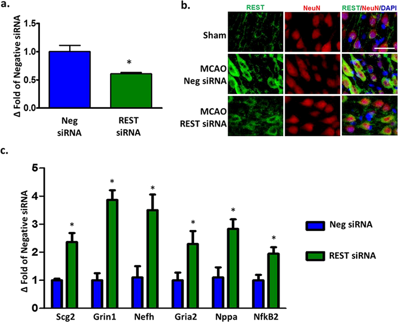
Fig. 2. REST knockdown de-repressed REST-responsive genes after focal ischemia.

Cortical REST expression at 24h of reperfusion following transient MCAO in siRNA and negative control siRNA groups (n=3/group) (a). Immunohistochemical staining showed co-localization of REST (green) with NeuN (red) and DAPI (blue) at 3 days of reperfusion (n=3/group). Scale bar = 30 μm (b). Real-time PCR of REST target genes at 24h of reperfusion (n=3/group) (c). Values are mean ± SEM. *p<0.05 versus negative siRNA.
