Fig. 1.
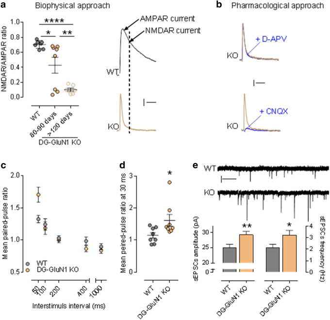
(a) Left panel, NMDAR/AMPAR ratio in cont and DG-GluN1 KO. AMPAR- and NMDAR-EPSC amplitudes are extracted from the dual component obtained at +40 mV, at 10 and 50 ms post-stimulus, respectively. Biophysical analysis of the dual component at +40 mV showed that NMDAR-mediated current in DG granular cells (dash line on the right panel) is absent in DG-GluN1 KO (4–5 months old). Note that the NAR decrease observed at 80–90 days is driven by 3/8 cells that were not exhibiting any NMDAR-mediated current (measured at 50 ms, dash line). Hash marks on left panel indicate group means ± SEM. (One-way ANOVA: F (2, 19) = 18.41: ****p < 0.0001; post hoc test: *p < 0.05; **p < 0.01). Calibration: 50 ms, 20 pA. (b) D-APV at 50 μM did not have any effect on evoked EPSC in DG-GluN1 KO (dual component obtained at 40 mV), indicating that NMDAR-mediated current is not present. AMPAR blockade with CNQX 10 μM almost totally eliminated evoked EPSC. Calibration: 50 ms, 20 pA. (c) Mean paired-pulse ratio values in CA3 pyramidal neurons from DG-GluN1 KO mice (n = 15 cells, 4 mice) is increased at an inter-stimulus interval of 50 ms compared to cont (n = 17 cells, 4 mice) (two-way ANOVA, Interaction PPR × inter-stimulus interval: F(4, 120) = 6.328, p = 0.0001; post hoc test at 50 ms: ***p < 0.01). (d) Mean paired-pulse ratio values in CA3 pyramidal neurons from DG-GluN1 KO mice (n = 8 cells, 3 mice) is also increased at 30 ms inter-stimulus intervals compared with neurons from cont (n = 8 cells, 3 mice) (t(14) = 2.294, *p < 0.05). (e) Top panel, sample traces of sEPSCs from neurons in cont and DG-GluN1 KO (KO) groups. Calibration: 1 s, 20 pA. Bottom panel, spontaneous EPSCs amplitude and frequency are increased in DG-GluN1 mice (n = 23 cells, 5 mice) compared with cont (n = 21 cells, 6 mice). Amplitude: t(42) = 2.819, **p = 0.007; frequency: t(42) = 2.194, *p = 0.034

