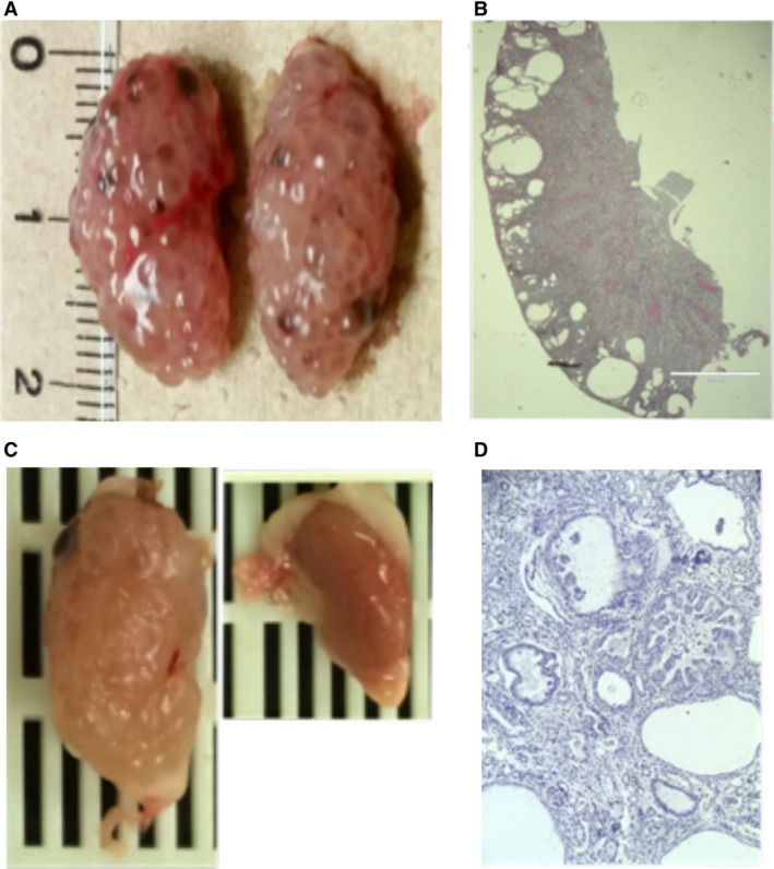
Figure 1.

Tsc renal cystic disease. (A) AqpCreTsc2 kidneys at 11 weeks with significant renal cystic disease. (B) Coronal sections of the kidney in figure A. (C) Mouse kidneys from RenCreTsc1 mouse with unilateral cystic disease. These are on the same size scale as in A. (D) Coronal section of kidney in figure C.
