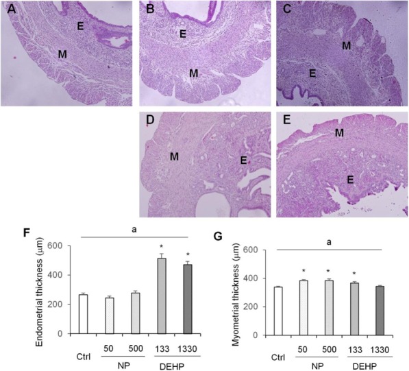
Fig. 3. Photomicrography of endometrium and myometrium and their thickness (μm) in response to NP and DEHP treatment. 10–12-week-old female mice were exposed to NP (50 or 500 μg/L) or DEHP (133 or 1,330 μg/L) in drinking water for 10 weeks. (A–E) Representative H&E stained uterus (×100). (A) Control. E, endometrium; M, myometrium, (B) NP 50 μg/L, (C) NP 500 μg/L, (D) DEHP 133 μg/L, (E) DEHP 1,330 μg/L, (F) Endometrium thickness (μm), DEHP 133 and 1,330 μg/L increased endometrium thickness, (G) Myometrium thickness (μm) including longitudinal and circular muscle layers. NP 50 and 500 μg/L and DEHP 133 μg/L increased myometrium thickness. Data are presented as means±SEM. a p<0.05, two way ANOVA; * p<0.05, significantly difference compared control vs. experimental group. Ctrl, control; NP, nonylphenol; DEHP, di-(2-ethylhexyl) phthalate.

